Reproduction and Egg Development (Experiment)
Alpheus parvirostris reproduce via sexual reproduction,with a male and female necessary for reproduction to be successful. Mating occurs when the male grabs the female and squeezes the muscles surrounding its ejaculatory duct, releasing a spermatophorcal cord into the female (Wicksten 2010). Fertilization occurs when the eggs pass through the endopods and pleomere sternite, collecting the sperm from the broken spermatophore (Wicksten 2010).
Alpheus parvirostris that were collected from Heron Island were always found in pairs (male and female), with the female supporting bright green eggs attached to the ovigerous setae located on her pleopods. Six pairs of shrimp were collected, with the females of all six bearing eggs. A sub-sample of the eggs were carefully removed from the females pleopods and fixed so that they would not degrade.
Spawning generally occurs shortly after the mating process, and is carried out as the eggs move out of the females gonopore and onto the pleopods (Wicksten 2010). Freshly spawned eggs contain large amounts of yolk (Wicksten 2010). The pleopods are critical to the eggs survival as the beat, providing fresh water and oxygen supplies to the eggs.
Timeline of Alpheus parvirostris egg development available eggs at time of collection. Here it is shown that initial eggs (newly produced) are made up of almost entirely of yolk, coloured green. Over time, the yolk supply diminishes as the larvae within the egg begins to develop. This can be seen in the second photo where the yolk supply is slowly being replaced by layers of cells. This is start of a process known as cleavage, where the yolk will eventually be surrounded by a single layer of cells (Wicksten 2010).
Cell division and embryonic development continues as seen in the final photo, where the majority of the yolk supply has been used and replaced by the outline of a developing shrimp. Note the presence of the eye (black spot) as well as the fine outline of the tail (left of egg).
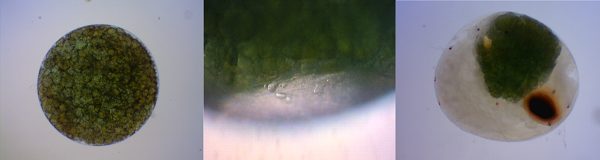 Timeline of Alpheus parvirostris egg development. Original Photo Aidan Janetzki 2013.
As time was limited on heron island, none of the collected eggs or eggs left on females hatched within the study period. However, below is a drawing of a generalized decapod larvae known as zoea. This illustrates a close representative of what newly hatched Alpheus parvirostris would look like, had they hatched. At this stage, zoea have two sessile eyes, two pairs of antennae, the mandibles, maxillipeds and pereopods (Wicksten 2010). It is believed to take between four and eleven larval stages fro the zoea to reach a juvenile stage (Wicksten 2010).

Drawing of generalized decapod larvae (zoea). Drawing by Aidan Janetzki 2013. Information adapted from Rupert et al. 2004.
To further examine the changes that occur during the Alpheus parvirostris egg life cycle, eggs of varying stages in development were stained with a fluorescence dye called DAPI. As DAPI binds strongly to A-T regions within DNA, it therefore highlights the nucleus of each cell because this is where DNA is stored within a cell. As an egg develops, cells divide and multiply, while at the same time using up the yolk supply. Thus, as the egg develops, an increase in cell (nucleus) should be observed. This phenomenon can clearly be seen below, where the difference in number of nuclei can be seen between eggs at different development stages. The left shows an undeveloped egg, where the majority of the egg is made up of a yolk supply. Note the limited amount of fluorescent nuclei. The middle and right photos show the same developed egg with varying strengths of fluorescence. Here, the outline of a developing shrimp can clearly be seen in the top half of both eggs. Cell concentration and therefore nuclei count is a lot higher in a further developed egg.
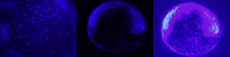
Timeline of Alpheus parvirostris egg development stained with DAPI under fluorescent light. Original Photo Aidan Janetzki 2013.
To further examine the changes in egg development, two eggs at different ages were section and stained. As seen in the DAPI dyed images, the further developed egg (right) clearly depicts the beginnings of juvenile shrimp. This is shown by the blue/purple aggregations in the egg, in the shape of a curled shrimp around the remaining yolk supply (pink). Alternatively, the relatively undeveloped egg (left) demonstrates the absence of any formation of shrimp, and is made up (almost) entirely of yolk.
 Timeline of Alpheus parvirostris egg development sectioned and stained. Viewed under a electron microscope. Original Photo Aidan Janetzki 2013.
Here we see a time-lapse video of Alpheus parvirostris eggs over one day. While this a relatively short period with respect to the entire development of the egg, we can still see cell movement and development particularly visible in the right egg at the base of the yolk supply. Here the yolk is being moved around and used up as the egg continues to develop.
Here we show a time-lapse video of Alpheus parvirostris eggs over twelve hours, with a focus on yolk migration. While studying the eggs under microscope, granules of yolk were observed to be travelling from one side of the egg to the other, in particular, from the tail of the developing shrimp to its head.
|